PET Imaging
Positron Emission Tomography (PET) is a medical imaging technology that allows physicians to visualize the body's abnormal cellular activity. PET scans produce digital pictures that can, in many cases, aid the physician in identifying several forms of cancer, damaged heart tissue and brain disorders.
A PET scan is very different from ultrasound, X-ray, MRI, or computerized tomography (CT) scans, which detect changes in the body structure or anatomy, such as a sizeable tumor or musculoskeletal injury. A PET scan can help physicians distinguish between living and dead tissue or between benign and malignant disorders, whereas other imaging technologies merely confirm the presence of a mass or abnormality.
Since a PET scan images the biology of disorders at the molecular level, it can help the physician detect irregularities in cellular activity at a very early stage, generally before anatomic changes are visible. A PET scan can, in many cases, help identify disease earlier and more specifically than ultrasound, X-ray, MRI or CT scans.
PET Utilization
The majority of PET scans are performed for oncologic applications. Physicians utilize PET scans for diagnosing, staging and evaluating treatments for their cancer patients. A PET scan helps the physician distinguish between living and dead tissue or between benign and malignant disorders, unlike other imaging technologies which merely confirm the presence of a mass. PET imaging provides the physician with additional information about cellular activity which guides the characterization of a questionable abnormality as malignant or benign.
A PET scan can show the extent of disease. For patients whose cancer is newly diagnosed, it is important to determine if the cancer has spread to other parts of the body so that appropriate treatment can be started. A PET scan images the entire body in a single examination, and aids the physician in detecting the primary site(s) as well as any metastases. Painful, costly and invasive surgery, such as thoracotomy, may no longer be necessary for diagnosis.
A PET scan can also help physicians monitor the treatment of disease. For example, chemotherapy leads to changes in cellular activity that is observable by PET imaging long before structural changes can be measured by ultrasound, X-ray, MRI or CT scans. This gives physicians an alternative technique to evaluate treatments earlier, perhaps even leading to modifications in treatment, before an evaluation would normally be made using other imaging technologies.
After treatment is complete, a PET scan allows the physician to investigate suspected recurrence of cancer, revealing tumors that might otherwise be obscured by scar tissue resulting from surgery and radiation therapy.
A PET scan puts time on your side. The earlier the diagnosis and the more accurate the assessment of the extent of disease, the better the chance for successful treatment.
Visualizing Disease
The PET scan begins with an injection of a glucose-based radiopharmaceutical, which travels through the body, eventually collecting in the organs and tissues targeted for examination.
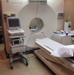
The patient lies flat on a table that moves incrementally through the PET scanner. The scanner has cameras that detect the gamma rays emitted from the patient, and turns those into electrical signals. These are processed by a computer to generate the images. The table moves slowly through the scanner and many sets of images are produced.
The acquired electrical signals are assembled by the computer into a 3-D image of the patient's body. If an area is cancerous, the signals will be stronger there than in the surrounding tissue, since more of the radiopharmaceutical will be absorbed in those areas.
PET is a highly sensitive procedure that aids in the detection of small cancerous tumors, and also subtle changes in the brain and heart. This enables physicians to identify and treat these diseases earlier and more accurately.