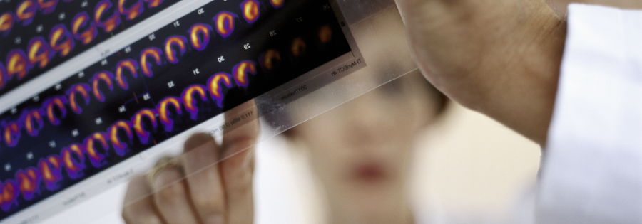
Cardiac PET
PET Scans and Heart Disease
PET imaging provides a way to assess the severity of heart disease and measure its impact on heart function. Clinical studies show an important role for PET in screening for coronary heart disease, assessing flow rates and flow reserves, and distinguishing viable from nonviable heart tissue. Two areas of clinical application have emerged:

Myocardial Perfusion
PET is used by physicians to reveal whether or not a patient has coronary artery disease, which is caused by accumulation of plaques within the walls of the arteries that supply blood to the heart.

Myocardial Viability
PET is used by physicians to determine the viability of heart muscle and if it has been permanently damaged due to decreased or absent blood flow.
Medicare Overview
The information below is taken from the Medicare National Coverage Determinations Manual:
The identification of patients with partial loss of heart muscle movement or hibernating myocardium is important in selecting candidates with compromised ventricular function to determine appropriateness for revascularization. Diagnostic tests such as 18F FDG PET distinguish between dysfunctional but viable myocardial tissue and scar tissue in order to affect management decisions in patients with ischemic cardiomyopathy and left ventricular dysfunction.
Medicare covers 18F FDG PET for the determination of myocardial viability as a primary or initial diagnostic study prior to revascularization or following an inconclusive SPECT.
Limitations: In the event a patient receives a SPECT test with inconclusive results, a PET scan may be covered. However, if a patient receives an 18F FDG PET study with inconclusive results, a follow up SPECT test is not covered.
Private Payer Overview
Private insurance typically follows Medicare coverage guidelines. However, private payers can have wide variation in coverage policies, coverage criteria or procedures. Additionally, most insurance companies require pre-authorization for a PET/CT scan. It is advisable to verify local specific coverage guidelines with the payer prior to performing the scan.